
The Eye as a Window to Cardiovascular Health: What the Retina Reveals About the Heart
The eyes have long been called the “window to the soul,” but they are also an extraordinary window into cardiovascular health.

The eyes have long been called the “window to the soul,” but they are also an extraordinary window into cardiovascular health. The retina provides a unique, non-invasive view of the body’s microvasculature—its small arteries, veins, and capillaries—where systemic diseases often manifest early. (1,2) Advances in retinal imaging and artificial intelligence (AI) now enable clinicians to detect cardiovascular disease (CVD) before symptoms appear, identify risk factors, and predict adverse events such as heart attacks and strokes. (3) This convergence of anatomy and technology is transforming routine eye exams into powerful tools for preventive medicine. (4)
The Biological Link: Why the Retina Mirrors the Heart
The connection between ocular and cardiovascular health lies in their shared vascular architecture. The retina is the only place in the body where arteries, veins, and capillaries can be directly visualized in vivo. (5) These vessels share similar structural and physiological properties with those in the heart and brain, making the eye a natural mirror of systemic vascular status. (6)
Damage to the vascular system—through endothelial dysfunction, altered shear stress from hypertension, neurovascular decoupling due to oxygen imbalance, or oxidative stress from ischemia—often appears first in the retina. (2) This has led to the rise of cardiac oculomics, the study of ocular biomarkers reflecting cardiovascular health.
Retinal findings currently associated with cardiovascular disease include:
- Narrowed arterioles and widened venules: classic signs of hypertension and heart failure.(7)
- Vascular lesions: retinal hemorrhages, microaneurysms, and cotton-wool spots indicating chronic vascular damage, often associated with hypertension or diabetes.(8)
- Reduced perfusion and capillary rarefaction: decreased vessel density revealing, microvascular dysfunction and chronic underperfusion.(9)
- Tortuous vessels: increased curvature or twisting of retinal arterioles or venules, reflecting vascular remodeling and hemodynamic stress associated with hypertension, atherosclerosis, and stroke. (10,11)
From Structure and Flow to Function: Technological Advances
Modern retinal imaging allows clinicians to move beyond visual observation, providing structural, circulatory, and increasingly, functional insights into vascular health.
Visualizing Structure and Blood Flow
Each imaging modality offers a unique perspective:
- Fundus Photography: Produces detailed two-dimensional images of the retinal surface, highlighting vascular changes such as hemorrhages, cotton-wool spots, and arteriovenous nicking—hallmarks of chronic hypertension. (12)
- Optical Coherence Tomography (OCT): Provides cross-sectional, microscopic images of retinal layers, enabling measurement of the Retinal Nerve Fiber Layer and detection of subtle ischemic lesions like Retinal Ischemic Perivascular Lesions (RIPLs). (13,14)
- Optical Coherence Tomography Angiography (OCT-A): Maps blood flow by tracking red blood cell movement without the need for dye injection, offering quantitative metrics such as vessel area density and foveal avascular zone size. (15)
Despite remarkable progress in retinal imaging, a critical gap remains: current modalities detect structural and circulatory changes only after significant metabolic disruption. It’s like seeing just the tip of an iceberg—fundus photography, OCT, and OCT-A capture visible damage, but the earliest metabolic alterations remain unseen. To move from reactive diagnosis to proactive prevention, clinicians must turn to functional biomarkers that reflect underlying metabolic activity. One technology promising to enable this shift is ocular spectroscopy.
Adding a Functional Perspective with Spectroscopy
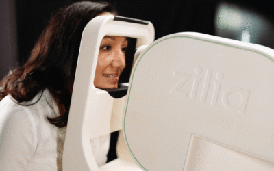
Ocular spectroscopy complements structural imaging by analyzing how light interacts with tissue to reveal its biochemical composition. Much like astronomers use starlight to infer the makeup of distant objects, ocular spectroscopy measures reflected light with unique absorption patterns characteristic of specific molecules within the eye.
The resulting spectral signature acts as a molecular fingerprint, revealing the presence of oxygenated and deoxygenated hemoglobin and other biomarkers. For example, ocular spectroscopy can quantify oxygen saturation in ocular tissues—providing early insight into metabolic dysfunction.(16)
Researchers are also exploring its potential to detect transthyretin amyloidosis, a systemic, underdiagnosed disease that can lead to heart failure.(17) Identifying its molecular markers non-invasively through the eye exemplifies the promise of retinal functional imaging for systemic disease detection.
The eye provides a sensitive, non-invasive means to monitor systemic vascular health and predict cardiovascular outcomes.
Connecting Retinal Biomarkers to Clinical Outcomes
An expanding body of research demonstrates that retinal signs can reflect cardiovascular disease severity and risk:
- Hypertension: Chronic high blood pressure produces narrowed arterioles, arteriovenous nicking, and decreased vessel density in both superficial and deep capillary layers on OCT-A. (18)
- Atherosclerosis and Coronary Artery Disease: Wider venules, cotton-wool spots, and subretinal drusenoid deposits correlate with coronary artery disease and myocardial infarction. Lower vessel density is linked to greater coronary artery stenosis.
- Stroke: A retinal artery occlusion (RAO) is now classified by the American Heart Association as an ischemic stroke equivalent, requiring urgent cardiovascular assessment. (19) Additional features—such as retinal hemorrhages and RIPLs—also correspond to increased stroke risk.
- Heart Failure: Wider venules predict a higher risk of heart failure and are associated with increased readmission and mortality. OCT-A studies frequently show reduced capillary density in these patients. (20)
Together, these findings confirm that the eye provides a sensitive, non-invasive means to monitor systemic vascular health and predict cardiovascular outcomes.
The Road Ahead: AI and Interdisciplinary Integration
Bringing retinal analytics into cardiovascular medicine presents great promise but also key challenges—chiefly the need for standardization, validation, and cross-disciplinary collaboration.
AI is accelerating discovery by detecting subtle vascular patterns beyond human perception. Deep learning models trained on large image datasets can estimate cardiovascular risk factors such as age, blood pressure, and smoking status from fundus images alone. (21) Yet these models rely on consistent imaging protocols and diverse, well-validated datasets to perform reliably across populations and devices.
Transparency remains a major hurdle, as AI systems often accurately predict outcomes but fail to explain their reasoning, limiting clinical adoption. Co-existing eye diseases can further confound algorithms, underscoring the need for careful data curation.
True progress will come from close cooperation among eye-care professionals, cardiologists, and data scientists. Each discipline brings a complementary perspective. As collaboration deepens, functional tools such as ocular spectroscopy will complement structural and flow-based imaging—enabling a comprehensive view of both vascular and metabolic health.
Conclusion: Redefining Systemic Health Through the Eye
The eye is more than an organ of sight—it is a living reflection of vascular and metabolic balance. Its transparent structures and accessible microvasculature make it uniquely suited for non-invasive exploration of systemic health.
The convergence of advanced imaging and functional spectroscopy marks a shift from population-level risk prediction to personalized, real-time evaluation of microvascular health. As interdisciplinary partnerships strengthen, the routine eye exam may soon take center stage in preventive cardiology. Ultimately, by looking into the eye, we may gain the clearest view yet of the heart.
References
- Jin K, Zhang J, Grzybowski A. Editorial: Predictive and diagnostic approaches for systemic disorders using ocular assessment. Front Med (Lausanne). 2024;11:1529861. Published 2024 Dec 11. doi:10.3389/fmed.2024.1529861
- Kellner RL, Harris A, Ciulla L, et al. The Eye as the Window to the Heart: Optical Coherence Tomography Angiography Biomarkers as Indicators of Cardiovascular Disease. J Clin Med. 2024;13(3):829. Published 2024 Jan 31. doi:10.3390/jcm13030829
- Bisen JB, Sikora H, Aneja A, Shah SJ, Mirza RG. Retinal Imaging as a Window into Cardiovascular Health: Towards Harnessing Retinal Analytics for Precision Cardiovascular Medicine. J Cardiovasc Dev Dis. 2025;12(6):230. doi:10.3390/jcdd12060230
- Meira Fogel-Levin S, Sadda SR, Rosenfeld PJ, et al. Advanced retinal imaging and applications for clinical practice: A consensus review. Surv Ophthalmol. 2022;67(5):1049-1078. doi:10.1016/j.survophthal.2022.02.005
- Lee SJV, Goh YQ, Rojas-Carabali W, et al. Association between retinal vessels caliber and systemic health: A comprehensive review. Surv Ophthalmol. 2025;70(2):184-199. doi:10.1016/j.survophthal.2024.11.009
- Egle M, Hamedani AG, Deal JA, et al. Retinal microstructure and microvasculature in association with brain amyloid burden. Brain Commun. 2025;7(1):fcaf013. doi:10.1093/braincomms/fcaf013
- Flammer J, Konieczka K, Bruno RM, et al. The eye and the heart. Eur Heart J. 2013;34(17):1270-1278. doi:10.1093/eurheartj/eht023
- Lim LS, Tan B, Ong YT, et al. Longitudinal evaluation of cotton wool spot following rapid glycemic control. Am J Ophthalmol Case Rep. 2024;33:102255. doi:10.1016/j.ajoc.2024.102255
- Zhou S, Yan Q, Guo J, He W, Zhao Y. Early diagnosis of coronary heart disease based on retinal microvascular parameters of OCTA. Front Cell Dev Biol. 2025;13:1654159. Published 2025 Aug 12. doi:10.3389/fcell.2025.1654159
- Poirier J, Allain G, Bansept MA, et al. Standardizing image acquisition and processing methods: A critical need for the accurate assessment of retinal blood vessel tortuosity. Biomed Opt Express. 2025;16(4):2739-2755. doi:10.1364/BOE.516004
- Ekhator C, Devi M, Barker C, et al. Arterial Tortuosity Syndrome: Unraveling a Rare Vascular Disorder. Cureus. 2023;15(9):e44906. doi:10.7759/cureus.44906
- Tripathy K, Modi P, Arsiwalla T. Hypertensive Retinopathy. In: StatPearls [Internet]. Treasure Island, FL: StatPearls Publishing; July 6, 2025. Available from: https://www.ncbi.nlm.nih.gov/books/NBK525980/
- Bellanda V, Delaney A, Schulgit MJ, et al. Screening for Retinal Ischemic Perivascular Lesions in Patients Undergoing Cardiovascular Assessment: A Cross-Sectional Study. Ophthalmol Retina. Published online 2025 Sep 9. doi:10.1016/j.oret.2025.09.002
- Madala S, Adabifirouzjaei F, Lando L, et al. Retinal Ischemic Perivascular Lesions, a Biomarker of Cardiovascular Disease. Ophthalmol Retina. 2022;6(9):865-867. doi:10.1016/j.oret.2022.05.005
- Untracht GR, Matos R, Dikaios N, et al. OCTAVA: An open-source toolbox for quantitative analysis of optical coherence tomography angiography images. arXiv. 2021. Available from: https://arxiv.org/abs/2109.01835
- Lapointe N, Akitegetse C, Poirier J, et al. Targeted Spectroscopy in the Eye Fundus. J Biomed Opt. 2023;28(12):126004. doi:10.1117/1.JBO.28.12.126004
- Zilia Inc. Zilia launches innovative project to detect a rare heart disease. Zilia Blog. Published 2024 May 15. Available from: https://ziliahealth.com/blog/ocular-spectroscopy-attr-amyloidosis-detection
- Lee WH, Park JH, Won Y, et al. Retinal microvascular change in hypertension as measured by optical coherence tomography angiography. Sci Rep. 2019;9(1):156. doi:10.1038/s41598-018-36474-1
- Mac Grory B, Schrag M, Biousse V, et al. Management of Central Retinal Artery Occlusion: A Scientific Statement From the American Heart Association. Stroke. 2021;52(6):e282-e294. doi:10.1161/STR.0000000000000366
- Bayat K, Pooyan P, Chhablani J, et al. Retinal and Choroidal Alterations in Heart Failure: A Systematic Review and Meta-analysis of OCT and OCT-A Findings with Emphasis on HFrEF. Ophthalmol Ther. 2025;14(11):2631-2651. doi:10.1007/s40123-025-01238-4
- Poplin R, Varadarajan AV, Blumer K, et al. Prediction of cardiovascular risk factors from retinal fundus photographs via deep learning. Nat Biomed Eng.2018;2(3):158-164. doi:10.1038/s41551-018-0195-0
Written by the Zilia Team on November 12, 2025
More on our Blog

Canadian Study on Retinal Oxygenation Using Zilia Ocular
Quebec City, February 20, 2026 – Zilia announced today that its Zilia Ocular device will be used in a new...
Zilia to Support Landmark Study on Retinal Degeneration and Regeneration
Quebec City, November 26, 2025 – Zilia, a health technology company at the forefront of oculomics, announced...
Zilia Launches Clinical Study to Investigate Ocular Oximetry as a New Glaucoma Biomarker
Quebec City, July 2, 2025 – Zilia is launching a new clinical trial in collaboration with Dr. Paul...

Solutions