
5 Best Reads in Ocular Oximetry
With all this buzz about ocular oximetry, where can you get more information on the subject? Rather than sifting through internet searches and databases, we’ve done the heavy lifting for you and here, we’ll give you five of the best resources on the topic.
Retinal Oximetry Discovers Novel Biomarkers in Retinal and Brain Diseases (1)
In this study, retinal oximetry is studied as a non-invasive method that can be used to diagnose early and stage several eye diseases such as diabetic retinopathy (DR), central retinal vein occlusion (CRVO), retinitis pigmentosa, glaucoma, as well as Alzheimer’s disease and Multiple Sclerosis.
DR and CRVO are conditions hallmarked by retinal hypoxia. When looking at oximetry in diabetic retinopathy, the authors found an increase in venous oxygen saturation and a decrease in arteriovenous difference. In both DR and CRVO, there is disturbed blood flow with ensuing tissue hypoxia, as well as production of vascular endothelial growth factor (VEGF). Both of these pathophysiological aspects cause changes in oxygen saturation. In both conditions, there is also a correlation between disease severity and the level of hypoxia.
The authors propose retinitis pigmentosa and glaucoma are both considered to be atrophic conditions since oxygen consumption is reduced and correlates with atrophy and reduction in visual field.
Lastly, the retina is an extension of the brain; with that, it is not surprising that we see ocular oximetry changes in Alzheimer’s and Multiple Sclerosis patients. While the potential of retinal oximetry seems clear, the authors conclude that there is a need to extend the number of prospective studies in this area.
Retinal oximetry in patients with ischaemic retinal diseases (2)
This literature review looked for associations between oxygen saturation and retinal ischemic disease. The authors have selected 10 publications of interest on the topic: 6 about DR and 4 on the topic of CRVO. In diabetes, all six studies revealed an association between increased retinal venous oxygen saturation and the presence of DR. They also found a correlation between the retinal venous oxygen saturation and the severity of DR. Most of the studies, four out of the six, also found an increase in retinal arterial oxygen saturation in DR. In the case of CRVO, all studies found an increase in the oxygen saturation of retinal veins, but not that of arteries.
This paper looked at studies using two-wavelength oximetry devices. Their number is limited by the fact that ocular oximetry is still a relatively new discipline. Furthermore, the only available literature involves cross-sectional studies or case series; there have not yet been any prospective studies or clinical trials on the subject. Nevertheless, the fact that there are measurable and predictable markers for oxygen saturation in these conditions lends promise to the use of ocular oximetry in the management of eye diseases.
Ocular oximetry would even reveal effects from hypoxia in systemic conditions such as heart and lung disease.
Retinal oximetry: Metabolic imaging for diseases of the retina and brain (3)
This study corroborates the utility of ocular oximetry in diseases of the retina and brain.
Diabetic Retinopathy (RD)
In DR, retinal venous oxygen saturation is elevated and the arteriovenous difference is progressively reduced as the condition advances. This could be explained by the production of VEGF following hypoxia. The article also reports that therapeutic interventions such as laser treatment and vitrectomy appear to be improving retinal oximetry.
Central Retinal Vein Occlusion (CRVO)
The severity of CRVO would be correlated with ocular oximetry findings. Specifically, increased peripheral ischemia, loss in visual acuity and augmented thickness of macular edema would be associated with poorer oxygen saturation in the retinal venules.
Glaucoma and Retinitis Pigmentosa
In both of these diseases, the retinal atrophy would be associated with reduced oxygen consumption. This would result in greater venous saturation and a reduction in the arteriovenous difference. In glaucoma, worse ocular oximetry values are seen with thinner optic disc rims, worse visual fields and reduced nerve fiber layer thickness. Similarly, in retinitis pigmentosa, retinal oxygen saturation would be affected with advanced atrophy and visual field deterioration.
Systemic Disease
Ocular oximetry would even reveal effects from hypoxia in systemic conditions such as heart and lung disease. Neurological conditions such as Alzheimer’s disease and Multiple Sclerosis have also shown a correlation with ocular oximetry values.
Retinal oximetry in glaucoma: investigations and findings reviewed (4)
We have long known that factors outside of intraocular pressure play into the pathophysiology of glaucoma. As such, disruption of blood flow to the optic nerve would be an important risk factor for the disease.
This article postulates that there could be one of two ways that blood circulation would be involved in glaucoma. The ‘vascular hypothesis’ suggests that the blood flow impairment is primary, causing resulting atrophy of the tissue. The ‘mechanical hypothesis’ proposes that non-vascular factors, such as increased intraocular pressure, would cause tissue atrophy. Oxygen demand would subsequently decrease.
Ocular oximetry could play a role in the study of these two non-mutually exclusive hypotheses. Mathematical models suggest that the mechanical hypothesis may be predominant in the pathophysiology of high tension glaucoma, while the vascular hypothesis may better explain normotensive glaucoma. Continued research in this area could help to identify new therapeutic targets in order to improve patient outcomes.
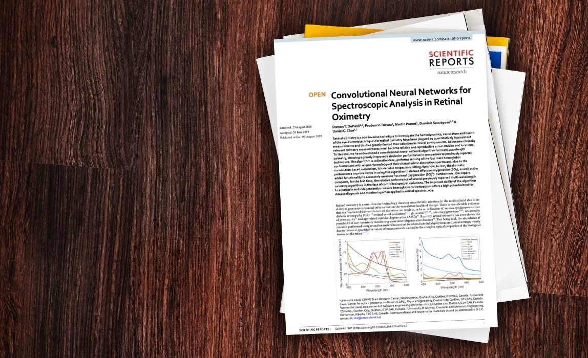
Convolutional Neural Networks for Spectroscopic Analysis in Retinal Oximetry (5)
This article aims to address the quantitative inconsistencies in ocular oximetry measurements, which has been limiting the clinical significance of this technology so far. The authors developed a convolutional neural network (CNN) algorithm for multi-wavelength oximetry. This calibration-free technique demonstrates the ability to sense the four different hemoglobin conformations and effectively deduce effective oxygenation and fractional oxygenation.
This method addresses three limitations that have historically plagued ocular oximetry measurement. First, First, the algorithms used in two or three wavelength systems are sensitive to scatter and absorption in the eye. Second, current systems often suffer from poor spectral calibration. Third, ocular oximetry has historically focused on effective oxygen saturation, while fractional oxygen saturation is more clinically relevant. The latter takes into account dyshemoglobins, which can affect the amount of circulating oxygen. The proposed CNN algorithm, due to its accuracy and repeatability, shows great promise for the clinical penetration of ocular oximetry.
Conclusion
There you have it, the five best reads in ocular oximetry. In short, ocular oximetry is a promising technology that could lead to earlier diagnosis and better management of many ocular, neurological and even systemic diseases. More work is needed to improve the accuracy and precision of these measurements. In this respect, Zilia’s new technology, Zilia Ocular, currently under development, is promising. This innovative technology involves the use of several wavelengths to obtain absolute and continuous oximetry measurements, opening the way to a wealth of metabolic information. This will certainly contribute to advancing research in this field and help protect the eyesight of many patients.
References
- Einar Stefánsson, Olof Birna Olafsdottir, Anna Bryndis Einarsdottir, Thorunn Scheving Eliasdottir, Thor Eysteinsson, Wouter Vehmeijer, Evelien Vandewalle, Toke Bek, Sveinn Hakon Hardarson; Retinal Oximetry Discovers Novel Biomarkers in Retinal and Brain Diseases. Invest. Ophthalmol. Vis. Sci. 2017;58(6):BIO227-BIO233. https://doi.org/10.1167/iovs.17-21776.
- Rilvén, S., Torp, T.L. and Grauslund, J. Retinal oximetry in patients with ischaemic retinal diseases. Acta Ophthalmol. 2017;95:119-127. https://doi.org/10.1111/aos.13229
- Stefánsson, E., Olafsdottir, O., Eliasdottir, T.S., Vehmeijer, W.B., & Hardarson, S. Retinal oximetry: Metabolic imaging for diseases of the retina and brain. Progress in Retinal and Eye Research. 2019;70:1-22.https://doi.org/10.1016/j.preteyeres.2019.04.001.
- Shughoury A, Mathew S, Arciero J, Wurster P, Adjei S, Ciulla T, Siesky B, Harris A. Retinal oximetry in glaucoma: investigations and findings reviewed. Acta Ophthalmol. 2020 Sep;98(6):559-571. https://doi.org/10.1111/aos.14397.
- DePaoli, D.T., Tossou, P., Parent, M. et al. Convolutional Neural Networks for Spectroscopic Analysis in Retinal Oximetry. Sci Rep. 2019; 9:11387. https://doi.org/10.1038/s41598-019-47621-7.
Written by the Zilia Team on March 5, 2021
More on our Blog
Zilia Ocular to Support Study in AMD and DME
Quebec City, March 9, 2026 – Zilia announced today that its Zilia Ocular device will be used in a new...
Canadian Study on Retinal Oxygenation Using Zilia Ocular
Quebec City, February 20, 2026 – Zilia announced today that its Zilia Ocular device will be used in a new...

Zilia to Support Landmark Study on Retinal Degeneration and Regeneration
Quebec City, November 26, 2025 – Zilia, a health technology company at the forefront of oculomics, announced...

Solutions