
Glaucoma Diagnosis and Management: What If We Could Intervene Sooner?
Glaucoma is a multifaceted ocular disease, which involves the degeneration of the optic nerve characterized by retinal ganglion cell loss.
Pathogenesis
Despite the ubiquity of glaucoma, our understanding of its pathogenesis remains incomplete. The primary risk factor for the disease is increased intraocular pressure (IOP), yet people with normal IOP can develop glaucoma too. In fact, normal tension glaucoma (NTG) comprises half of glaucoma cases in the United States of America; in Japan, it accounts for up to 90% of cases. This reinforces the fact that IOP is not the only etiology for optic nerve compromise and that, very often, glaucoma progresses despite intensive IOP-lowering treatments. We now know that many factors influence the development of glaucoma, including hereditary, vascular, immune, and structural elements. (1)
Diagnostic Delay
Glaucoma is often asymptomatic in its early stages. For this reason, among others, it can be challenging to detect. (1) One study found a diagnostic delay in 43% of cases of glaucoma. A quarter of cases had a total delay ranging from 3-27 years. (3) This is concerning because there is potential for significant vision loss over this time frame. Earlier treatment can postpone or even prevent visual disability during one’s lifetime. (3) Low socioeconomic status plays a role in diagnostic delays, as individuals may not have access to the resources necessary for detection. However, individual factors are not the only influences at play. Failures at the healthcare provider level also serve a role in diagnostic delay, through inaccuracy of case detection and/or delayed referral. (4)
A growing number of studies show that physiological changes such as dysregulation of blood flow and oxygenation take place long before irreversible damage occurs.
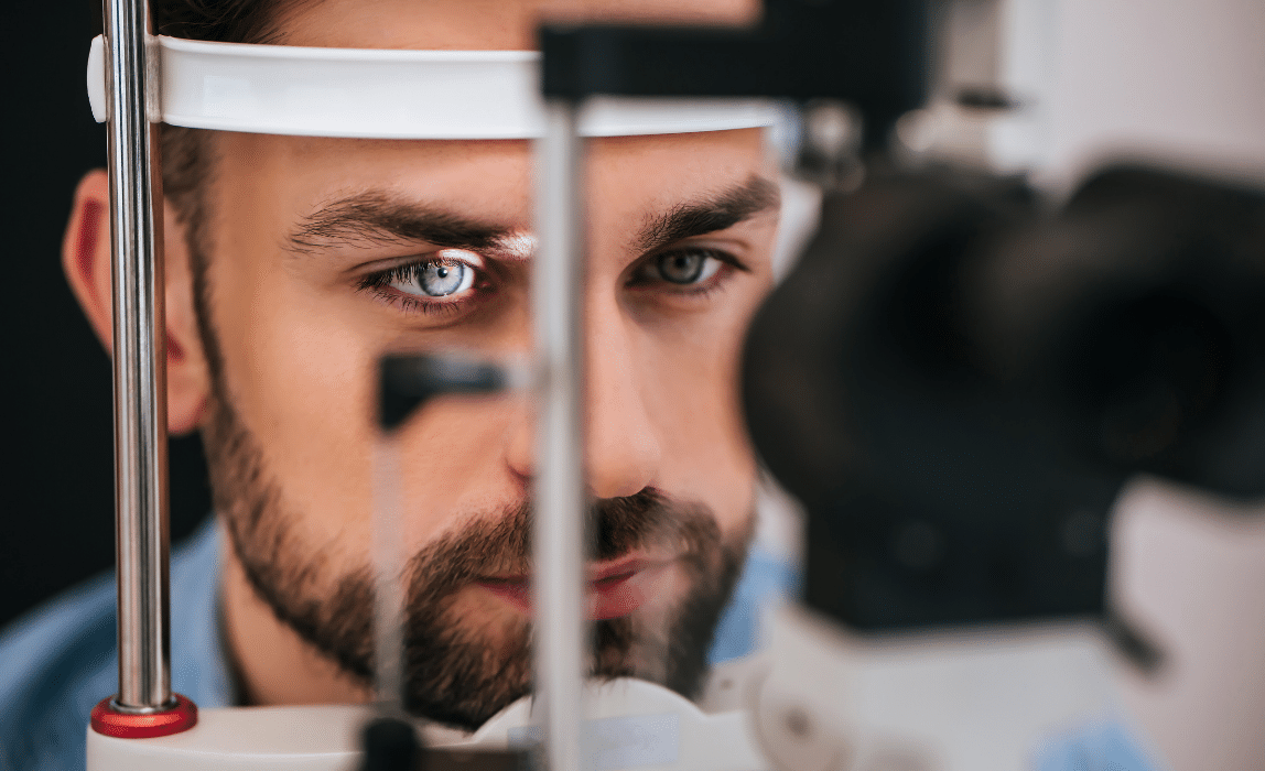
Diagnostic Tools
To understand how glaucoma diagnosis and management can be improved, let’s take a look at how we screen for glaucoma today.
Visual Field Testing
Visual field testing looks for functional loss of vision. Several metrics can be examined to evaluate the extent of visual field loss. The Mean Deviation (MD) gives a weighted average of field loss across the entire visual field. It does this by comparing the overall sensitivity to that of age-matched controls. (5) The Pattern Standard Deviation (PSD) also uses age-matched controls, but examines the difference in sensitivity between adjacent points. (6) Other metrics may be used as well, such as the Visual Field Index (VFI) and the Glaucoma Progression Analysis (GPA).
While visual field testing is an excellent way to measure vision loss late in the disease progression, glaucoma needs to be diagnosed before this functional impairment manifests.
Retinal Photography
Photographing the optic nerve can help to visualize changes in its structure over time. The degeneration of the optic nerve in glaucoma causes cupping, which can be measured by the cup-to-disc ratio. (7) An increasing ratio over time signals progressive loss of retinal ganglion cell axons. Photography can help to visualize rim thinning and notching, cup-to-disc enlargement or asymmetry, disc hemorrhaging, or nerve fiber layer defects, all of which raise suspicion for glaucoma. (8)
While fundus photography is helpful in documenting changes in the optic nerve over time, these nuances tend to be subtle and therefore difficult to visualize over short periods of time. Furthermore, these deviations cannot be quantified with photography. By the time a change is visible, the structural loss is significant.
Optical Coherence Tomography
Optical Coherence Tomography (OCT) is another tool used to monitor for glaucoma. This technology quantifies the disc and macular topography, as well as the nerve fiber layer (NFL) and ganglion cell complex (GCC) thickness around the optic nerve and the macula. (8) Monitoring with OCT helps to identify a change in these layers over time.
Although OCT is a useful tool in screening for glaucoma, it does have some limitations. The demarcation of the optic nerve may be inaccurate in some cases. (9) Certain conditions also make it especially difficult for the machine to produce accurate and useful data, such as high myopia, tilted disc, or extensive peripapillary atrophy. Furthermore, OCT measures structural loss, which is still unfortunately irreversible.
Future Technologies
What if we could find evidence of impending glaucoma before there’s been any structural loss, let alone a functional decline? If we could identify the loss of metabolic function responsible for the eventual demise of optic nerve tissue, perhaps we could intervene sooner?
A growing number of studies show that physiological changes such as dysregulation of blood flow and oxygenation take place long before irreversible damage occurs. (10)

Ocular Oximetry
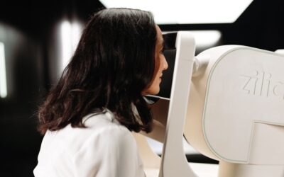
Ocular oximetry measurement allows for the monitoring of metabolic function over time, potentially enabling earlier diagnosis of ocular diseases such as glaucoma. This technology could also serve to evaluate the metabolic response to better inform treatment over time.
Zilia Ocular is the first retinal camera offering the continuous measurement of oxygenation in targeted regions of the eye. This device combines imaging and oximetry for the non-invasive measurement of metabolic function.
The use of ocular oximetry could revolutionize the way we approach glaucoma screening, diagnosis and treatment worldwide. By identifying the condition before structural and functional damage ensues, innovative therapies could intercept the pathophysiology of the disease at an earlier stage in the process. Ongoing research investigates which therapies would best address oxygen dysregulation. So far, there is evidence that carbonic anhydrase inhibitors, magnesium, and calcium-channel blockers may be beneficial in targeting this pathway. (11) We know that lowering IOP doesn’t address all the factors that play a role in glaucoma; new drug targets are needed to more comprehensively treat this disease.
Conclusion
The increasing incidence of glaucoma, and the delay in its diagnosis, is a public health concern. Zilia Ocular may serve to intercept glaucoma before it has a chance to ravage the structure and function of the eye. By identifying metabolic dysfunction sooner, treatment could potentially be initiated preemptively, using current and/or innovative therapies. The earlier identification and treatment of glaucoma have significant potential to prevent vision loss, ultimately improving the quality of millions of lives worldwide.
References
- Allison K, Patel D, Alabi O. Epidemiology of Glaucoma: The Past, Present, and Predictions for the Future. Cureus. 2020;12(11):e11686. Published 2020 Nov 24. doi:10.7759/cureus.11686
- Baneke AJ, Aubry J, Viswanathan AC et al.The role of intracranial pressure in glaucoma and therapeutic implications. Eye. 2020;34:178–191. doi:10.1038/s41433-019-0681-y
- Eissa IM, Abu Hussein NB, Habib AE, El Sayed YM. Examining Delay Intervals in the Diagnosis and Treatment of Primary Open Angle Glaucoma in an Egyptian Population and Its Impact on Lifestyle. J Ophthalmol. 2016;2016:7012826. doi:10.1155/2016/7012826
- Prior M, Francis JJ, Azuara-Blanco A, Anand N, Burr JM; Glaucoma screening Platform Study group. Why do people present late with advanced glaucoma? A qualitative interview study. Br J Ophthalmol. 2013;97(12):1574-1578. doi:10.1136/bjophthalmol-2013-303813
- Saunders LJ, Russell RA, Kirwan JF, McNaught AI, Crabb DP. Examining visual field loss in patients in glaucoma clinics during their predicted remaining lifetime. Invest Ophthalmol Vis Sci. 2014;55(1):102-109. Published 2014 Jan 7. doi:10.1167/iovs.13-13006
- Heo DW, Kim KN, Lee MW, Lee SB, Kim CS. Properties of pattern standard deviation in open-angle glaucoma patients with hemi-optic neuropathy and bi-optic neuropathy. PLoS One. 2017;12(3):e0171960. Published 2017 Mar 1. doi:10.1371/journal.pone.0171960
- Weinreb RN, Aung T, Medeiros FA. The pathophysiology and treatment of glaucoma: a review. JAMA. 2014;311(18):1901-1911. doi:10.1001/jama.2014.3192
- An G, Omodaka K, Hashimoto K, et al. Glaucoma Diagnosis with Machine Learning Based on Optical Coherence Tomography and Color Fundus Images. J Healthc Eng. 2019;2019:4061313. Published 2019 Feb 18. doi:10.1155/2019/4061313
- Sathyan P, Shilpa S, Anitha A. Optical Coherence Tomography in Glaucoma. J Curr Glaucoma Pract. 2012;6(1):1-5. doi:10.5005/jp-journals-10008-1099
- Wentz SM, Kim NJ, Wang J, Amireskandari A, Siesky B, Harris A. Novel therapies for open-angle glaucoma. F1000Prime Rep. 2014;6:102. Published 2014 Nov 4. doi:10.12703/P6-102
- Mozaffarieh M, Flammer J. Is there more to glaucoma treatment than lowering IOP?. Surv Ophthalmol. 2007;52 Suppl 2:S174-S179. doi:10.1016/j.survophthal.2007.08.013
Written by the Zilia Team on September 16, 2021
More on our Blog
Zilia Ocular to Support Study in AMD and DME
Quebec City, March 9, 2026 – Zilia announced today that its Zilia Ocular device will be used in a new...
Canadian Study on Retinal Oxygenation Using Zilia Ocular
Quebec City, February 20, 2026 – Zilia announced today that its Zilia Ocular device will be used in a new...
Zilia to Support Landmark Study on Retinal Degeneration and Regeneration
Quebec City, November 26, 2025 – Zilia, a health technology company at the forefront of oculomics, announced...

Solutions