
The Eye as Window to Brain Pathology
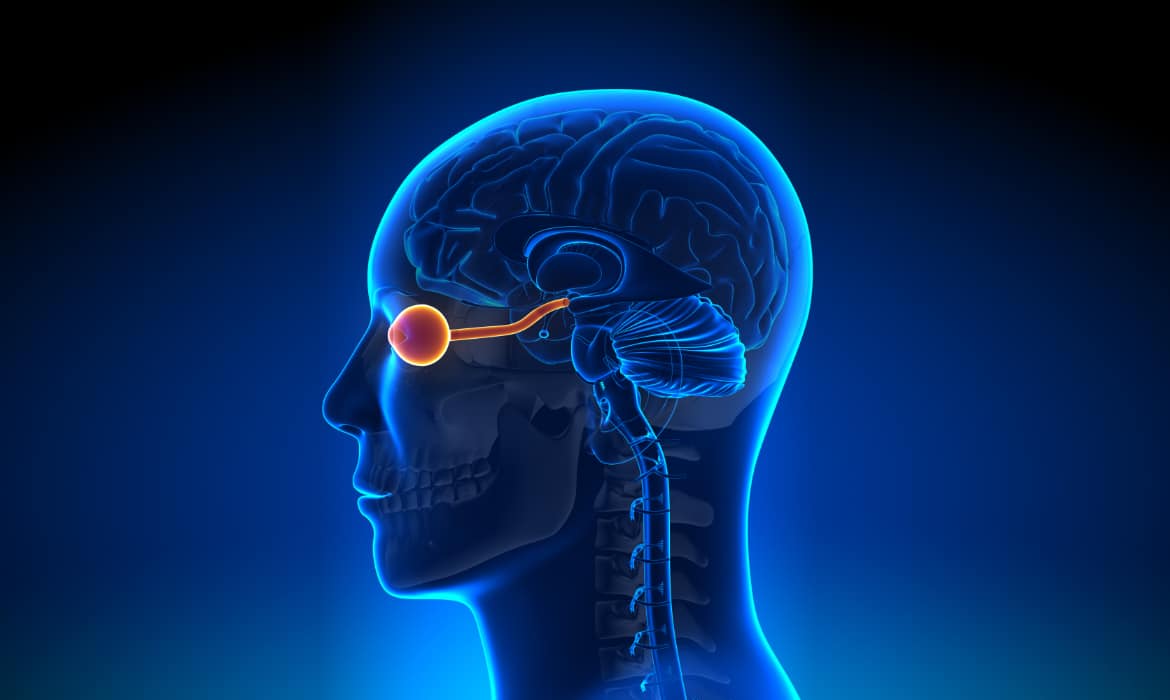
The eyes may be windows to our soul and psyche, but they are also windows to our health. In embryonic development, the optic nerve and retina extend from the developing brain. These structures are considered part of the central nervous system.
The eyes are the only organs in the human body where the central nervous system and blood vasculature are visible. The eye’s transparent media allows for visualization of the retina in vivo using noninvasive measures. Additionally, many disease processes affect the eye before changes occur in the rest of the body. Using modern imaging techniques, these unique features of the eye can be used for early detection of systemic disorders.
Link to the Brain
Growing evidence indicates that biomarkers of neurodegenerative diseases are found in the eye. These novel biomarkers have important implications for neurological conditions such as Alzheimer’s disease, Parkinson’s disease, and multiple sclerosis. Thus, the neurosensory retina may truly be a window into the pathologies of the brain.
Alzheimer’s disease, the most common form of dementia, can be challenging to diagnose in its early stages. The symptoms are primarily nonspecific and difficult to distinguish from normal, age-related changes (1). However, we know today that metabolic changes happen years before symptoms manifest (2). Retinal biomarkers offer a potential method for early detection and diagnosis of these changes and can have a sizable impact on the socioeconomic burden of these diseases. The potential benefits to both patients and healthcare systems are significant.
To better understand underlying vascular mechanisms of brain diseases, several studies have turned their focus on oxygen saturation.
Vascular Component
Vascular dysfunction contributes to the pathogenesis of many ocular diseases, such as glaucoma, diabetic retinopathy, hypertensive retinopathy, and age-related macular degeneration (3, 4). For example, stiffening of retinal arteries affects venous blood flow (particularly at arteriovenous crossing points), increasing the risk for retinal vein occlusions. Patients with diabetes, hypertension, or high cholesterol are particularly at risk. Similar retinal venous changes are suspected in Parkinson’s disease (3). These changes may preclude functional damage and help to identify patients with preclinical disease.
Neurovascular coupling, also called functional hyperemia, describes the relationship between neuronal activity and changes in cerebral blood flow. In the eye, visual stimulation (such as from a flicker of light) of the retina results in increased neuronal activity, leading to increased blood flow to the retina and optic nerve (5). Thus, neurovascular coupling allows the body to maintain the metabolic demands of neural tissue.
Although the mechanisms of neurovascular coupling are not fully understood, evidence suggests that dysfunction of this system occurs in various neurodegenerative and vascular diseases. Research shows that neurovascular coupling may be affected in Alzheimer’s disease, in which the retinal vessel response to light is reduced (6). Other studies suggest that similar events occur in multiple sclerosis, although further research is needed to support this theory (7). The vascular response to visual stimuli shows promise as a noninvasive method to diagnose and monitor neurological disorders.
Oxygen Saturation
To better understand underlying vascular mechanisms of brain diseases, several studies have turned their focus on an important biomarker of many ocular and systemic diseases: oxygen saturation. Oximetry measures the oxygen saturation of hemoglobin, while ocular oximetry measures blood oxygen saturation in the retina. Similarly to the brain, the retina has a high metabolic demand (8). Impairment of blood flow and oxygen supply to the retina can lead to pathological changes. Ocular oximetry can detect these changes in the retinal oxygen metabolism in both ocular and systemic diseases.
Studies find that the retina displays elevated oxygen saturation in Alzheimer’s disease (9). In multiple sclerosis, ocular oximetry findings show increased venular oxygen saturation, which indicates reduced oxygen uptake. Researchers believe that the reduced demand for oxygen may be related to atrophy following optic neuritis in multiple sclerosis patients (10).
Conclusion
Blood flow and oxygen play crucial roles in the maintenance of homeostasis. Current evidence shows that alterations in vascular regulation and oxygen saturation can lead to various pathologies of the central nervous system. Retinal biomarkers allow for the early detection of disease. However, there is a demand for technology to provide objective assessments of these biomarkers.
Traditional ocular oximeters measure blood oxygen saturation in large blood vessels and do not allow measurement of the retinal microvasculature. They only capture a single qualitative measurement. Zilia Ocular, on the other hand, uses the entire spectrum of light to measure oxygen saturation in the eye. This new technology platform offers continuous, quantitative measurements which provide more comprehensive insights on the oxygenation of the retina.
More studies are needed to validate the role of oxygen saturation and vascular dysfunction in neurological diseases, and more metabolic biomarkers have yet to be discovered, but the potential is tremendous. Novel technologies, such as Zilia Ocular, have the capacity to revolutionize the diagnosis and management of neurodegenerative disorders, including Alzheimer’s disease, multiple sclerosis, and Parkinson’s disease. With an aging global population, ocular oximetry will become increasingly important in clinical and research settings.
References
- Yap TE, Balendra SI, Almonte MT, Cordeiro MF. Retinal correlates of neurological disorders. Ther Adv Chronic Dis. 2019;10:2040622319882205. Published 2019 Dec 2. doi:10.1177/2040622319882205
- Morris JC. Early-stage and preclinical Alzheimer disease. Alzheimer Dis Assoc Disord. 2005;19(3):163-165. doi:10.1097/01.wad.0000184005.22611.cc
- Kromer R, Buhmann C, Hidding U, et al. Evaluation of retinal vessel morphology in patients with Parkinson’s disease using optical coherence tomography. PLoS One. 2016;11(8):e0161136. Published 2016 Aug 15. doi:10.1371/journal.pone.0161136
- Boeckaert J, Vandewalle E, Stalmans I. Oximetry: recent insights into retinal vasopathies and glaucoma. Bull Soc Belge Ophtalmol. 2012;(319):75-83
- Garhöfer G, Chua J, Tan B, Wong D, Schmidl D, Schmetterer L. Retinal neurovascular coupling in diabetes. J Clin Med. 2020;9(9):2829. Published 2020 Sep 1. doi:10.3390/jcm9092829
- Kotliar K, Hauser C, Ortner M, et al. Altered neurovascular coupling as measured by optical imaging: a biomarker for Alzheimer’s disease. Sci Rep. 2017;7(1):12906. Published 2017 Oct 10. doi:10.1038/s41598-017-13349-5
- Stickland R, Allen M, Magazzini L, Singh KD, Wise RG, Tomassini V. Neurovascular coupling during visual stimulation in multiple sclerosis: A MEG-fMRI study. Neuroscience. 2019;403:54-69. doi:10.1016/j.neuroscience.2018.03.018
- Country MW. Retinal metabolism: A comparative look at energetics in the retina. Brain Res. 2017;1672:50-57. doi:10.1016/j.brainres.2017.07.025
- Stefánsson E, Olafsdottir OB, Eliasdottir TS, et al. Retinal oximetry: Metabolic imaging for diseases of the retina and brain. Prog Retin Eye Res. 2019;70:1-22. doi:10.1016/j.preteyeres.2019.04.001
- Einarsdottir AB, Hardarson SH, Kristjansdottir JV, Bragason DT, Snaedal J, Stefánsson E. Retinal oximetry imaging in Alzheimer’s disease. J Alzheimers Dis. 2016;49(1):79-83. doi:10.3233/JAD-150457
- Einarsdottir AB, Olafsdottir OB, Hjaltason H, Hardarson SH. Retinal oximetry is affected in multiple sclerosis. Acta Ophthalmol. 2018;96(5):528-530. doi:10.1111/aos.13682
Written by the Zilia Team on April 20, 2021
More on our Blog

Zilia Ocular to Support Study in AMD and DME
Quebec City, March 9, 2026 – Zilia announced today that its Zilia Ocular device will be used in a new...

Canadian Study on Retinal Oxygenation Using Zilia Ocular
Quebec City, February 20, 2026 – Zilia announced today that its Zilia Ocular device will be used in a new...

Zilia to Support Landmark Study on Retinal Degeneration and Regeneration
Quebec City, November 26, 2025 – Zilia, a health technology company at the forefront of oculomics, announced...

Solutions