Vascular Dysregulation and Ocular Disease
Nearly 4.2 million Americans aged 40 and older suffer from uncorrectable vision impairment (1). As the population ages, this number is expected to grow significantly. Glaucoma, diabetic retinopathy (DR), and age-related macular degeneration (AMD) are the leading causes of vision loss.

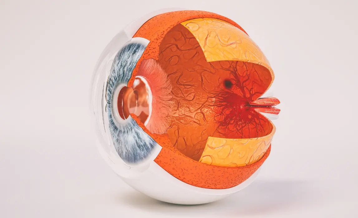
Current clinical tools such as retinal photography and optical coherence tomography (OCT) focus on assessing existing damage to the eye. However, a growing number of studies show that physiological changes such as dysregulation of blood flow and oxygenation take place long before irreversible damage occurs.
Diabetic Retinopathy
DR is a serious complication of diabetes that affects approximately 50% of those with Type 2 disease. In industrialized countries, it is the leading cause of blindness among those under the age of 65 (2).
Excess sugar in the blood progressively weakens the capillary wall. This degradation manifests itself in leaks, and eventually, in the rupture of tiny retinal vessels responsible for the exchange of gases and nutrients between cells and the blood.
Over time, large areas of the retina lose perfusion. In response, the retina produces new very fragile vessels. This is called neovascularization. Untreated, this phenomenon can progress and extend to the macula, where the center of the vision is located. As a result, the macula thickens (macular edema) and visual acuity decreases. In addition, these fragile new vessels can bleed into the vitreous, causing fibrosis. This can lead to retinal detachment, which can have devastating visual consequences.
Because DR is linked to vascular disturbances, the measurement of oxygen supply in the eye may serve as both a diagnostic marker and a tool to monitor response to therapy (3). Ocular oximetry may noninvasively improve early diagnosis and risk stratification in patients with diabetes.
Recent research has shown that ocular oximetry could become an important clinical tool in glaucoma, potentially enabling earlier diagnosis and treatment
Glaucoma
Glaucoma is an eye disease that causes permanent damage to the visual field, starting peripherally. This decrease in vision is the result of damage to the optic nerve, which enables the communication between the eye and the brain. More than three million Americans ages 40 and older are living with glaucoma. The vast majority of these people (90%) are affected by the most common form, open-angle glaucoma (OAG) (4).
It is often unknown why glaucoma develops. While heredity seems to be a risk factor, the scientific community is increasingly united in evoking a vascular cause to the observed damage to the optic nerve. Indeed, there is consensus regarding the effect of intraocular pressure combined with a lack of perfusion, and thus a lack of oxygen supply to the optic nerve head. As a result, precious ganglion cells gradually die.
There is accumulating evidence that dysregulation of oxygen in the eye is a major pathophysiological factor of OAG. Recent research has shown that ocular oximetry could become an important clinical tool in glaucoma, potentially enabling earlier diagnosis and treatment (3).
Age-Related Macular Degeneration
The macula is located in the center of the retina and provides detailed central vision. It is used to read, drive, and recognize faces. AMD blurs central vision while leaving peripheral vision intact. It is the leading cause of blindness in North America among those 55 and older (5).

The causes appear to be multiple and difficult to identify in a particular individual. Major risk factors include heredity, smoking, high blood pressure, and prolonged exposure to UV light. There are two forms of this disease: dry AMD, which accounts for 85 % of cases, and wet AMD which accounts for about 15 % of cases. Although there is currently no curative treatment for dry AMD, nutritional supplements are recommended to reduce the risk of progression. On the other hand, there are treatments for wet AMD, in order to preserve the visual acuity of patients.
The disease is often asymptomatic in the early years and regularly goes unnoticed during routine examinations. Recent studies have demonstrated a correlation between the decrease in choroidal circulatory parameters and the severity of AMD, pointing towards a potential role for ischemia in AMD (6). Since ischemic regions are poorly vascularized, new oximetry technologies could play an important role in further understanding of how oxygenation affects the pathophysiology of AMD (7).
Conclusion
It is increasingly recognized that eye diseases such as glaucoma, diabetic retinopathy, and age-related macular degeneration have a vascular component. It has been shown that oxygen dysregulation often precedes structural damage. Thus, in order to improve the understanding of ocular metabolism and facilitate its implementation in eye care, there is a need for accurate ocular oximetry measurement that can be performed in real-time and in all of the fundus tissues. With such a direct assessment of the vascular health of the retina, it would be possible to predict which patients are at risk for developing the diseases in question and to put in place adequate treatments before damage occurs.
References
- Centers for Disease Control and Prevention, Fast Facts of Common Eye Disorders. https://www.cdc.gov/visionhealth/basics/ced/fastfacts.htm. Published June 9, 2020. Accessed October 22, 2020.
- Fédération Française des Diabétiques, La rétinopathie diabétique et les maladies des yeux. Accessed October 22, 2020. https://www.federationdesdiabetiques.org/information/complications-diabete/retinopathie
- J. Boeckaert, E. Vandewalle, and I. Stalmans, “Oximetry: recent insights into retinal vasopathies and glaucoma,” Bull Soc Belge Ophtalmol, vol. 319, pp. 75–83, 2012.
- Bright Focus Foundation, Glaucoma: Facts and Figures. https://www.brightfocus.org/glaucoma/article/glaucoma-facts-figures. Published June 27, 2019. Accessed October 22, 2020.
- Foundation Fighting Blindness. What is Age-Related Macular Degeneration? https://www.fightingblindness.org/diseases/age-related-macular-degeneration. Accessed October 22, 2020.
- J. E. Grunwald, T. I. Metelitsina, J. C. DuPont, G.-S. Ying, and M. G. Maguire, “Reduced Foveolar Choroidal Blood Flow in Eyes with Increasing AMD Severity,” Invest. Ophthalmol. Vis. Sci.,vol. 46, no. 3, pp. 1033–1038, Mar. 2005.
- A. Geirsdottir, S. H. Hardarson, O. B. Olafsdottir, and E. Stefánsson, “Retinal oxygen metabolism in exudative age-related macular degeneration,” Acta Ophthalmol. (Copenh.), vol. 92, no. 1, pp. 27–33,Feb. 2014.
Written by Dr. Dominique Meyer MD, FRCSC, MBA on December 3, 2020
More on our Blog
Zilia Partners with Kagawa University for Groundbreaking Retinal Oxygenation Study in Japan
Quebec City, Apr. 30, 2024 - Zilia, a pioneer in non-invasive ocular biomarker technologies, announces a...
Oxygen’s Complex Role in Retinopathy of Prematurity
Premature birth is a remarkable feat of medicine. However, this early entrance into the world can sometimes...
Oxidative Stress and Eye Health
Oxygen plays an important role in our universe, being the third most abundant element and the second most...
Solutions


